Eye Diseases of Griffons-2016 Health Seminar

Melbourne EyeVet
Dr Anu O’Reilly & Dr Chloë Hardman P: 1800 EYE VET
W: www.melbourneeyevet.com.au E: [email protected]
Griffon Bruxellois
Eye Diseases known or suspected to be inherited
-
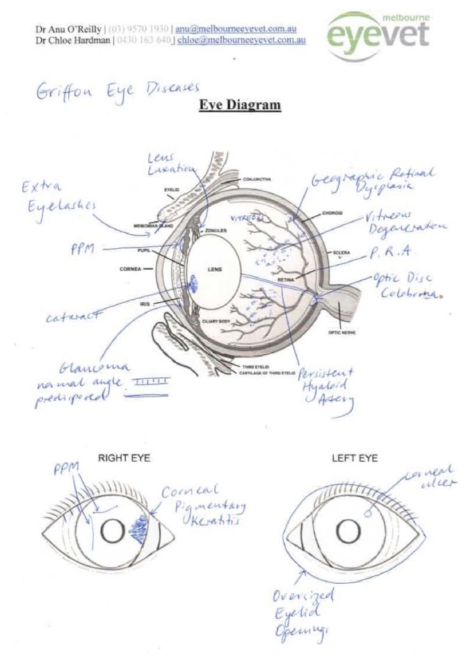
- Extra eyelashes
-
- Oversized eyelid openings – macroblepharon/exposure keratopathy syndrome
-
- PPM – persistent pupillary membrane
-
- Cataract
-
- Lens luxation
-
- Persistent hyaloid artery
-
- Vitreous degeneration
-
- Retinal atrophy – generalised
-
- Retinal dysplasia – geographic
-
- Optic nerve coloboma
-
- Glaucoma
-
- Corneal ulcers
Extra eyelashes/Distichia: these are eyelashes growing right on the edge of the eyelid from the openings of eyelid glands. 2-3% of US CERF examinations have distichiasis. If fine and in low numbers, they are unlikely to cause irritation. Surgery is required if they are causing clinical signs, including squinting, watery discharge and corneal ulcers.
Oversized eyelid openings: this is apparent in dogs with scleral show i.e. the white sclera is visible when the dog is alert. This can lead to exposure and drying of the cornea, corneal ulcers and scarring. Breeding dogs with severe
PPM (persistent pupillary membrane): PPM are usually strands of iris tissue that did not dissolve completely during the formation of the eye. They can be fine or thick and can run from the iris to iris, lens or the cornea. Iris to iris strands that are fine are of no concern, however iris to lens or iris to cornea strands result in an opacity at the attachment site. Breeding not advised.
Cataract: these are opacities in the lens. The lens should be crystal clear for perfect vision. With age, the lens will develop a cloudy/blue appearance but this needs to be differentiated from cataracts. Age-related change just causes problems with focusing. Any cataract in a young animal should be considered to be inherited and breeding is not advised.
Lens Luxation: This occurs when the zonules holding the lens either degenerate or break. Many terrier breeds are predisposed to this. The lens either falls backwards or comes forward and blocks drainage in the eye resulting in glaucoma. This is painful and surgery is required to relieve pain. If diagnosed early, lens removal surgery may be performed to try to save vision in the eye. Dogs affected with this condition are not suitable for breeding.
Persistent Hyaloid Artery: The hyaloid artery supplies blood to the lens during development of the eye. Once the eye is fully formed, blood flow in this artery stops and it is resorbed (fades away). If this process is not complete, an eye may have a persistent hyaloid artery. This can be related to more severe developmental problems. A dog affected with this is unlikely to have issues (although there is a chance that the vessel could bleed) however breeding is not advised.
Vitreous degeneration: The vitreous is the jelly at the back of the eye. Sometimes this goes through premature ageing and opacities and floaters can develop. In severe cases, vision is affected. Furthermore, the once firm vitreous that used to support the lens liquefies (changes from a thick gel to a watery substance), then lens loses its support and zonules may then break, leading to lens luxation.
Retinal atrophy (generalised): This condition is known as PRA – progressive retinal atrophy and is seen in many breeds. It is an inherited condition whereby the retina (nerve tissue) undergoes degeneration/thinning and results in vision loss. Initially there is night blindness, followed by day blindness and later cataracts. Affected dogs may be blind as young as 2 years old, however the more common age for blindness is 5-8 years old. Breeding is not advised, however with DNA testing, affected dogs may be bred to clear dogs to create a whole litter of carrier dogs.
Geographic retinal dysplasia: Many breeds may be affected with this condition. It is a more severe form of multi- focal retinal dysplasia, in which folds are seen in the retina. In these cases, there are multiple folds that line up to form a ring. The retina within the ring is predisposed to retinal detachment. Breeding is most definitely not advised.
Optic disc coloboma: This is also a developmental condition whereby the optic nerve head has not formed properly and there is missing nerve tissue in the form of a hole. These can also predispose the eye to retinal detachment and large holes can affect vision. Breeding not advised.
Glaucoma: Primary glaucoma develops as a result of a malformed drainage angle. The normal drainage angle allows easy flow of the fluid produced inside the eye (aqueous) into the drainage angle, and into the bloodstream. If normal flow holes are not formed, at some point the drainage becomes compromised, and the intraocular pressure increases. This is glaucoma, and high pressure in the eye is painful and causes vision loss by stopping blood flow to the nerve tissue (optic nerve and retina).
Other eye conditions seen in Griffons – breed predisposition
Corneal ulcers: A corneal ulcer occurs when there is a breach in the surface of the eye = cornea. They are usually caused by trauma (e.g. scratch) but are more common in dogs with oversized eyelid openings, dry eye, extra eyelashes and ectopic cilia (ingrown eyelashes). Most corneal ulcers when treated early can be resolved. Surgery is required in some cases if the cause if due to an eyelash or the eyelid openings are enormous and recurrent ulcers are occurring. Tear readings should always be measured once the ulcers have healed to rule out dry eye as an underlying cause.
Pigmentary keratitis: This condition is extremely common in Pugs with 50% of Pugs having some degree of pigment develop on the cornea. It is also seen in Griffons. It is usually due to eyelid conformation (oversize or entropion = inward rolling eyelids). Treatment includes Tacrolimus drops, and or surgery to burr the pigment off and corrective eyelid surgery.
Eye certification in Griffons
As a general rule, in a breed with very few recorded problems, eye certification may be performed at 1-2 years of age (prior to first breeding). This will enable examination for extra eyelashes, eyelid size and conformation, persistent pupillary membrane, persistent hyaloid artery, geographic retinal dysplasia, optic disc coloboma and predisposition to glaucoma (gonioscopy). It will also examine for cataract, lens laxity and retinal atrophy, however these may still develop at a later age.
A check around 4-5 years of age and a final check around 8 years of age (if clear this will clear the dog of inherited eye disease) will be able to give breeders a clear picture of what their breeding stock is developing.
AVA ACES results - Summary
|
No presented |
% of annual registrations |
Schedule 1 |
Schedule 2 |
Repeat defects |
litters |
|
|
2009 |
7 (6 nor) |
7.22 |
0 |
HC |
0 |
0 |
|
2010 |
4 (1 nor) |
2.53 |
0 |
0 |
3 i-i PPM |
0 |
|
2011 |
12 (10 nor) |
8.45 |
0 |
0 |
PHA, cat |
0 |
|
2012 |
1 (1 nor) |
0.5 |
0 |
0 |
0 |
0 |
|
2013 |
15 (14 nor) |
10.1 |
0 |
0 |
1lip 1 HR |
0 |
|
2014 |
6 (6 nor) |
3.6 |
0 |
0 |
0 |
0 |
|
2015 |
6 (6 nor) |
3.0 |
0 |
0 |
0 |
0 |
Average number of registrations = 97 + 158 + 142 + 200 + 151 + 166 + 200/7 = 159.1 per year
2009: Griffon Bruxellois (7 presented, 7.22% of annual registrations) nil Sch 1, HC is Sch 2, nil repeat defects, nil litters (Sch 1, Sch 2, repeat defects, litters)
-
2010: Griffon Bruxellois (4, 2.53%) nil nil PPM’s (I-I) 3 nil (1 of 4 showed no lesions)
-
2011: Griffon Bruxellois (12, 8.45%) nil nil pers. hyaloid+cat 1, PRA + sec. cataract 1 nil (10 of 12 showed no lesions)
-
2012: Griffon Bruxellois (1, 0.5%) nil nil nil No litters 1 showed no lesions Gonioscopy done on 1 – normal
-
2013: Griffon Bruxellois (15, 10.1%) nil nil corn lipid 1 hyaloid remnant 1 0 (14 of 15 normal)
-
2014: Griffon Bruxellois 6 total and 6 unaffected. 6 adults is 3.6% of annual registrations. While numbers are down
on last year, the apparent lack of evidence of a major threat to vision is encouraging. More useful data could be produced with sample sizes >10%.
2015: Griffon Bruxellois 2 6 total and 6 unaffected. 6 adults is 3.0% of annual registrations. While numbers are still fairly low, the apparent lack of evidence of a major threat to vision is encouraging. More useful data could be produced with sample size >10%
CERF results – eye certification results from USA
These results are published in the ‘Blue Book’ (see references). This is available as a download and found online when you search for ‘Blue Book’.
A summary of the findings for the past 25 years follows. The main conditions seen at CERF exams were:
-
Extra eyelashes 1.7-2.7% prevalence
-
Pigmentary keratitis 1.2-6.6% prevalence
-
PPM 2.8-13.2% prevalence
-
Cataract 3.7-7.5% with anterior cortical most commonly seen (front edge of lens)
-
Vitreous degeneration 14.6-27.1%
-
Retinal folds up to 4%
-
PRA 1.7-2.7%
-
Optic disc coloboma 1.0-2.5%
Genetic Testing of inherited disorders
There are currently no health-related DNA tests available for the Brussels Griffon in Australia, UK or the USA. In the near future tests should be available for PRA and syringomyelia.
General eye health – how to tell if your dogs’ eyes are healthy
-
- Menace response – responds to movement towards its face
-
- Bright and shiny – suggests good tear production
-
- White of the eyes white
-
- Minimal to no discharge – small amount of mucoid discharge can be normal
-
- Torch examination – pupil response, clarity of front of the eye, retinal reflection and lens examination
References
Ocular Disorders presumed to be inherited in purebred dogs. 7th Edition, 2014. Genetics Committee of the American College of Veterinary Ophthalmologists http://www.acvo.org/new/diplomates/resources/BlueBook2014-7thEdition.pdf
Australian Veterinary Association ACES (Australian Canine Eye Scheme)
http://www.ava.com.au/aces
-
Contact Details
President: Mrs Colleen De Haan [email protected]
Secretary -Mrs Robin Simpson [email protected]
Puppy enquiries - Beth Canavan [email protected]
0490085215
